Cystic liver tumours can be classified as simple liver cysts, cystadenomas, or parasitic echinococcal cysts. Simple liver cysts, which originate from small bile ducts, can occur singly, multiply or as part of polycystic liver disease, predominantly affecting women.
Although the exact cause of simple cysts is unknown, there are suggestions that they may be congenital or linked to the use of oral contraceptives.
In general, simple cysts do not require specific treatment, as most patients do not experience any symptoms, and they do not usually develop into cancer. Therefore, monitoring at regular intervals is recommended. Treatment is only necessary if the cyst cause discomfort. Cystic liver tumours typically cause discomfort or pain under the right rib cage, and localization in the left lobe of the liver can lead to stomach irritation, nausea, and vomiting after meals.
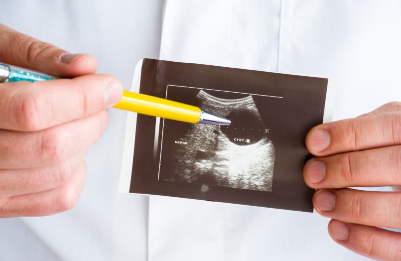
With the increasing use of ultrasound, scanners, and magnetic resonance imaging, cystic changes in the liver have become an increasingly common finding. However, accurate diagnosis may be challenging, and a biopsy may be required to confirm the nature of the cystic lesion.
Cyst sclerosing is an intervention in which a solution is injected through a needle into the cyst cavity, guided by ultrasound and performed by an interventional radiologist. This approach is preferable to surgery, which is reserved for cases where cyst sclerosing has failed. Biliary cystadenomas must be completely removed, as they have the potential for malignant transformation. Laparoscopic surgery is the preferred approach.